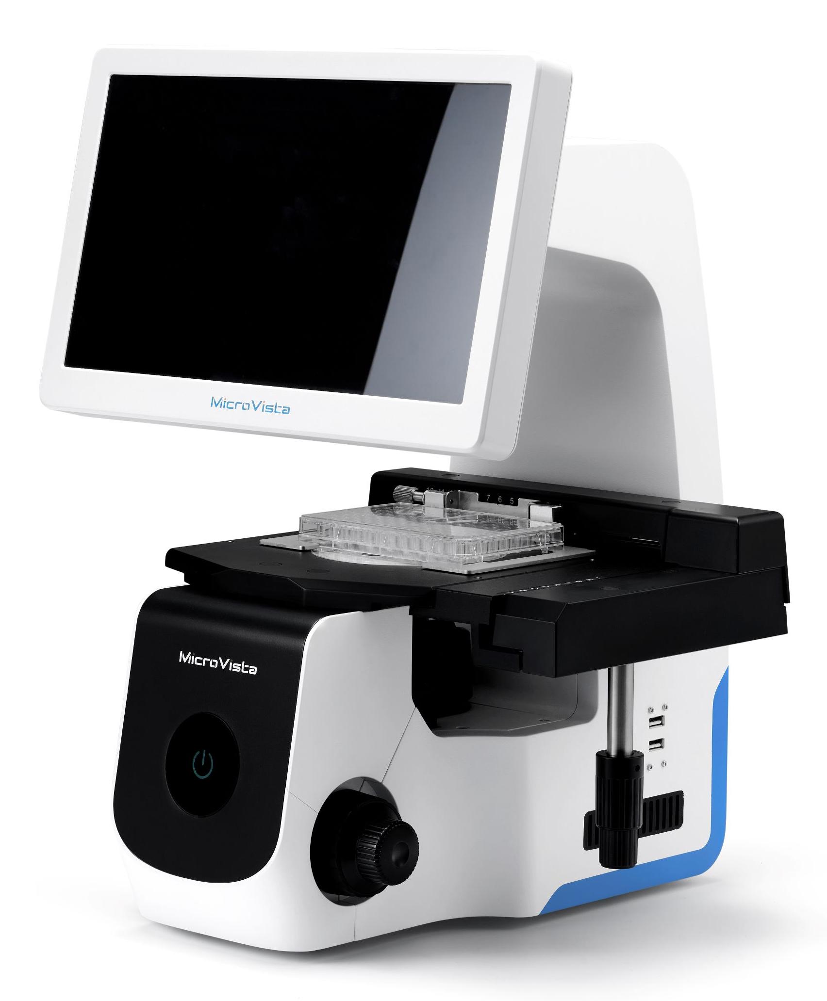
Premium imaging and effortless operation with Excellent optical design
Vita-X1 is an innovative All-in-One imaging system. As an eyepiece-free microscope,it not only guarantees exceptional image resolution with consistently, but also significantly streamlines workflows through the intuitive interface. Vita-X1 could provides satisfactory performance in various applicaiton environment,such as cell-culture check, cell sampling and processing, live cell monitoring, tissue observation,etc.,making it an ideal tool for life science research.
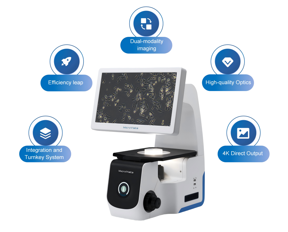
01/Integration and Turnkey System
Vita-X1 adopts a highly integrated design. Its all-in-one body significantly saves laboratory space, making it particularly suitable for compact environments with limited space. It's also compatible with biosafety cabinets or clean bench, ensuring the sterility of samples for some experiments with high�level cleanliness requirements,such as stem cell culture and cell therapy etc.
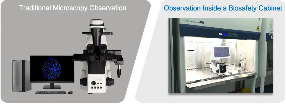
Isolateing external contamination to ensure
the sterility of experimental samples
02/Efficiency leap
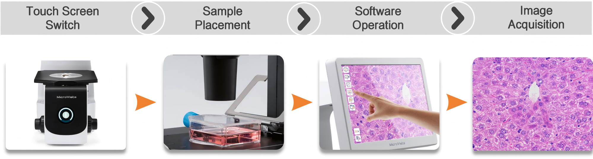
The integrated and intuitive design of Vita-X1 significantly improve experimental efficiency. Only one button could switch on or off all the imaging units including illumination light source, camera, and HD display, etc.. The user could operate whole system with a gentle touch on the screen or a mouse click. one-click on the image capture icon could simultaneously conduct image acquisition and automatic storage on an external drive. Video recording is also possible to capture fast dynamic moments.
Workflow:
03/Dual-modality imaging
Modular design offers various imaging methods with flexibility.
Vita-X1 is configured with both brightfield and phase contrast imaging modes, providing flexible and efficient observation solutions for different sample types:
| 
|
Observation of the open field |
Phase difference observation |
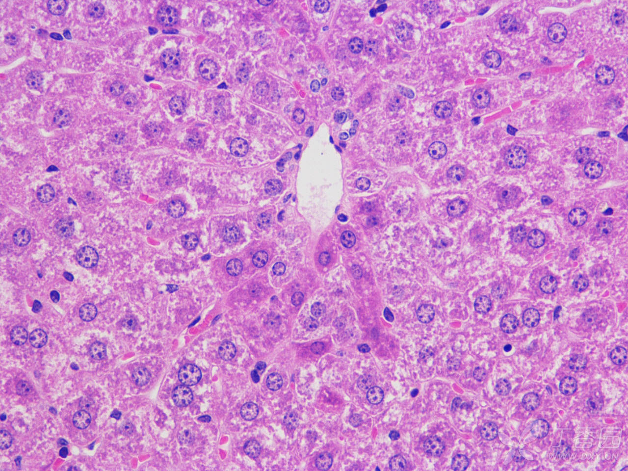
Brightfield observation is suitable for stained or high-contrast samples (e.g., tissue sections, stained cells) with simple operation and low cost.
| Phase contrast observation is designed for transparent or unstained live samples (e.g., live cells, microorganisms), enhancing the contrast without staining.
|
04/High-quality Optics
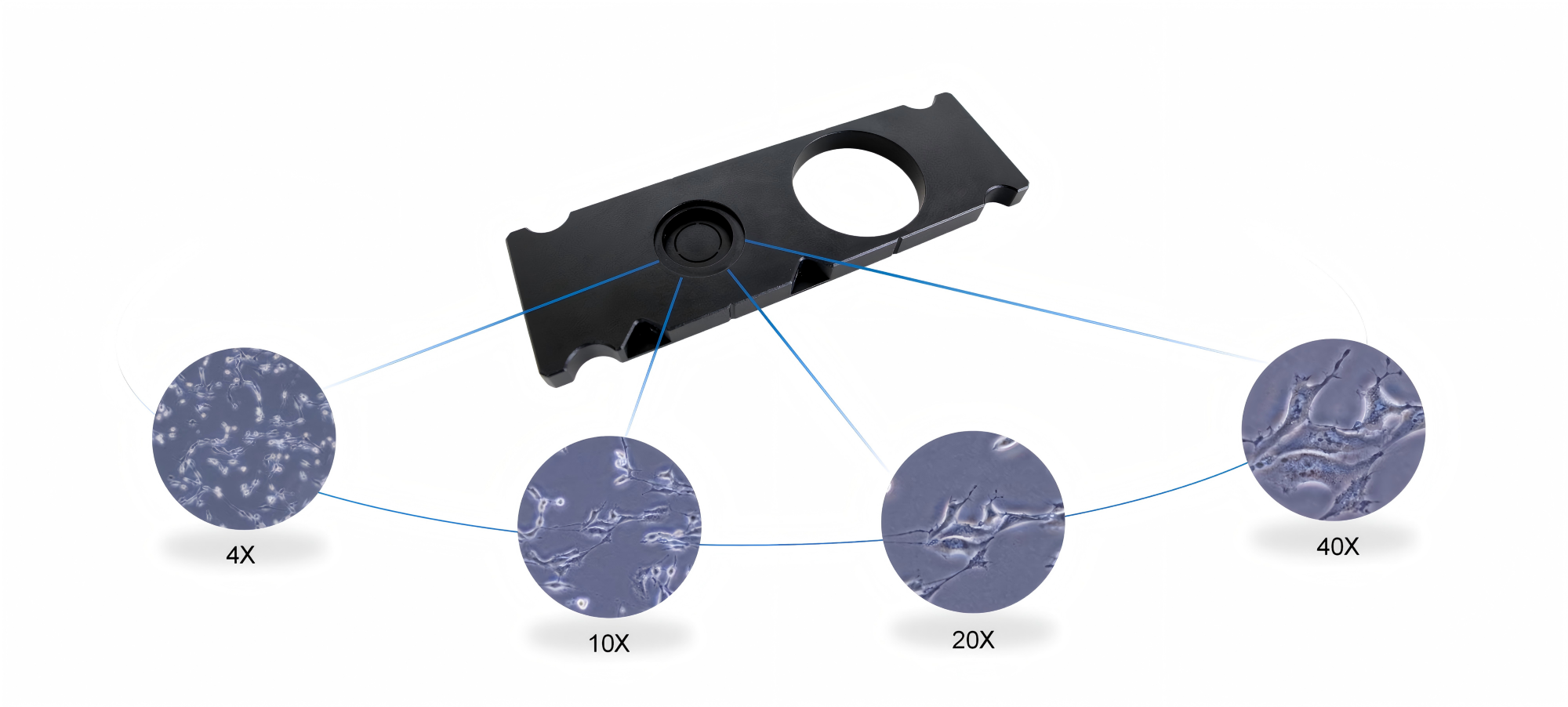
World-class top Optical Quality | 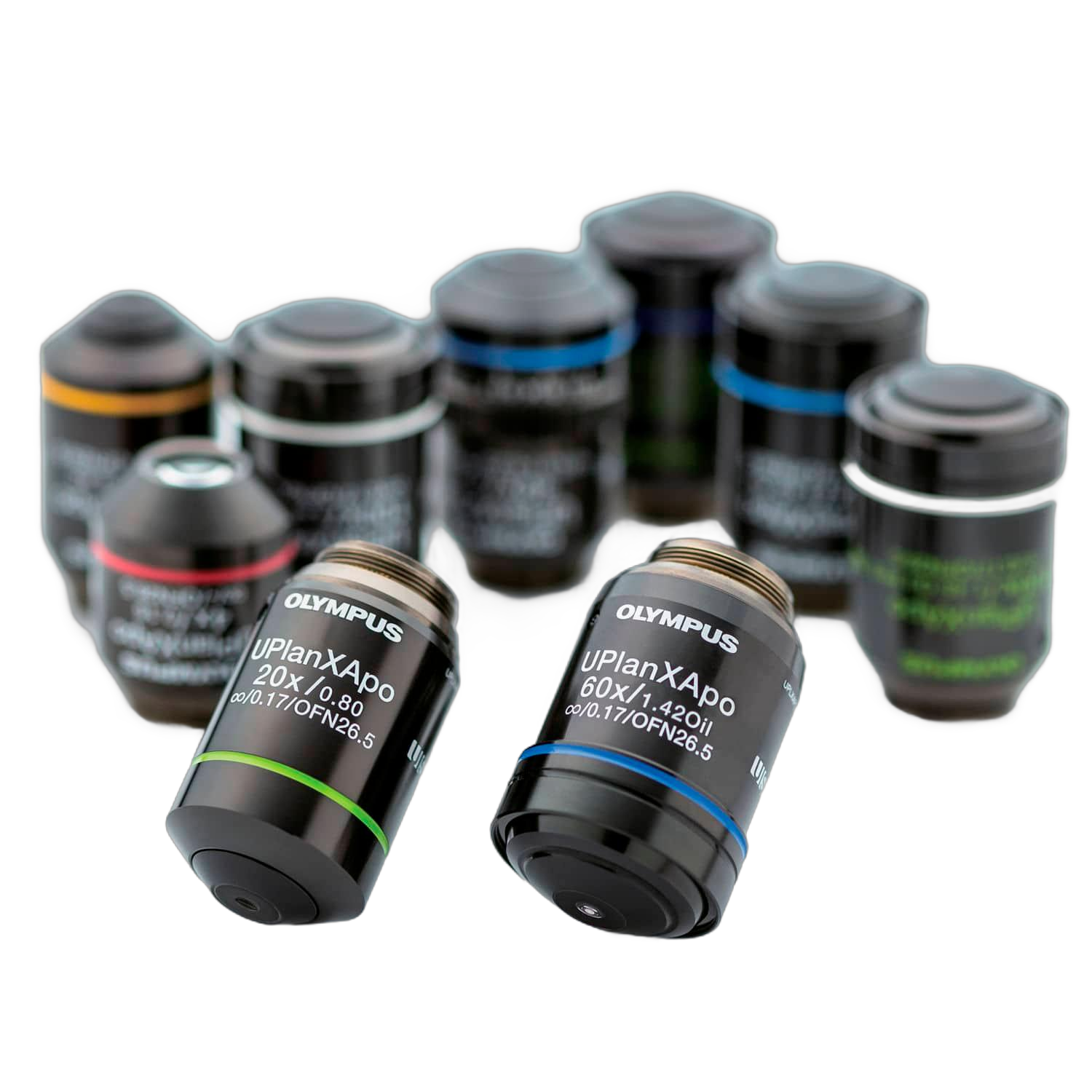 | Six High-Precision Objective for option |
the world-class objectives delivers high-resolution, high contrast images to ensure clear visibility of fine structures in cells and tissues. | Objective with the magnification of 4X, 10X, 20X, 40X, 60X, 100X, covering the full scale from macroscopic samples to microstructural observation.
|
|
|
Superior Phase Contrast Imaging
| Intelligent Optical Path Adjustment |
Only one phase ring is required for different magnifications (4X-40X). Ensures excellent phase contrast imaging while improving observation efficiency.
| Köhler illumination system with automatic calibration ensures uniform illumination across different magnifications, avoiding glare or shadow interference.
|
LED Light Source with ultra-Long Working-time
The high-performance LED illumination system boasts a working time over 20,000 hours. Combined with optimized optical design of microscope, it ensures bright and uniform illumination across the entire field of view. Its excellent fidelity also accurately presents the authentic colors of samples. The LED light source could significantly reduces energy consumption while providing a consistently stable illumination, greatly enhancing the efficiency and reliability of prolonged microscopic observation. | 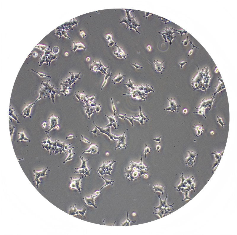 |
|
Clear observation under the entire field of vision |
High-contrast phase contrast observation |
Compatible with various culture vessels
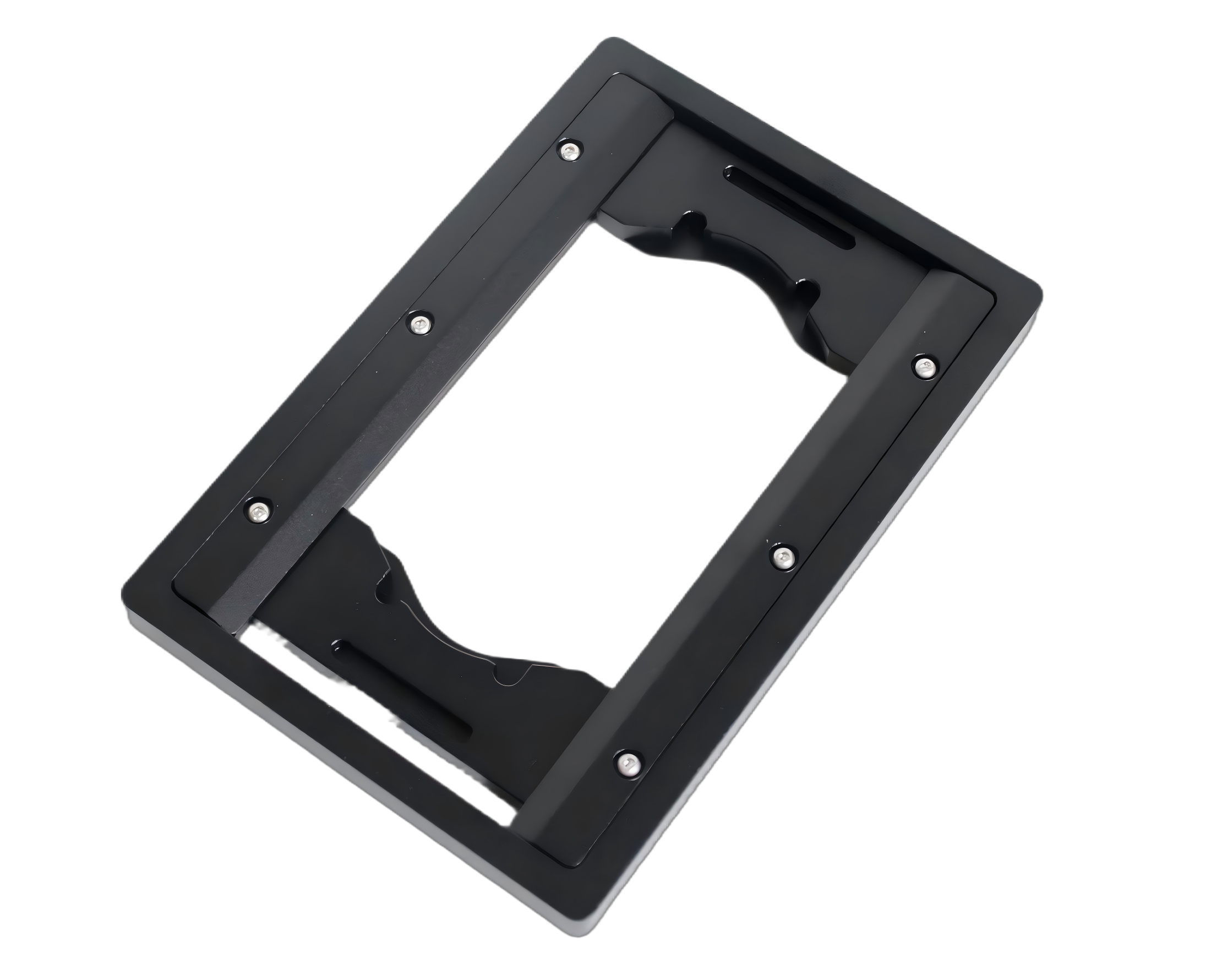
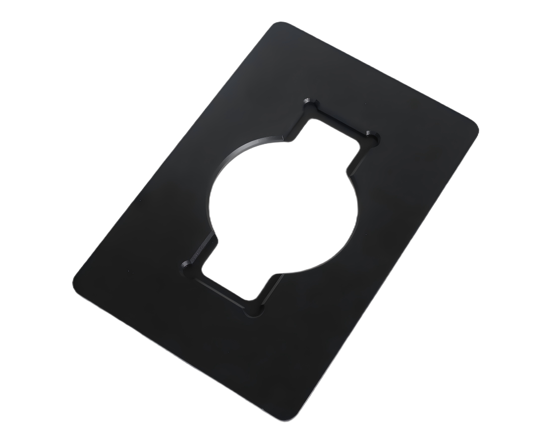
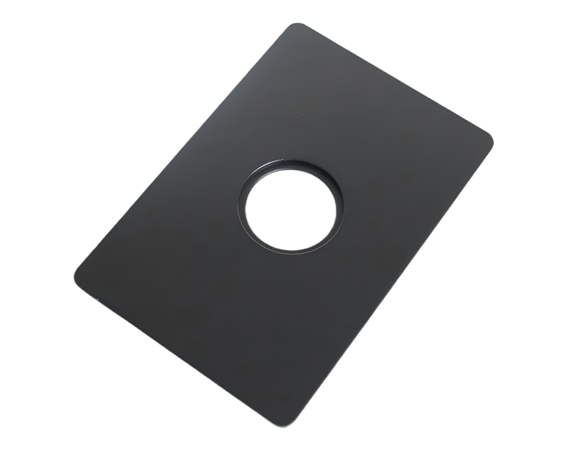
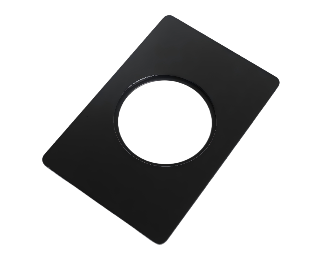
Vita-X1 is equipped with multiple adapters (Universal, dish-type and slide-type adapters), which allows easily observation of specimens in various cell culture containers for diverse experimental.
 | X1-HOUN | X1-HOS |
| 
|
X1-HOD35 | X1-HOD60 |
| 
|
05/4K Direct Output
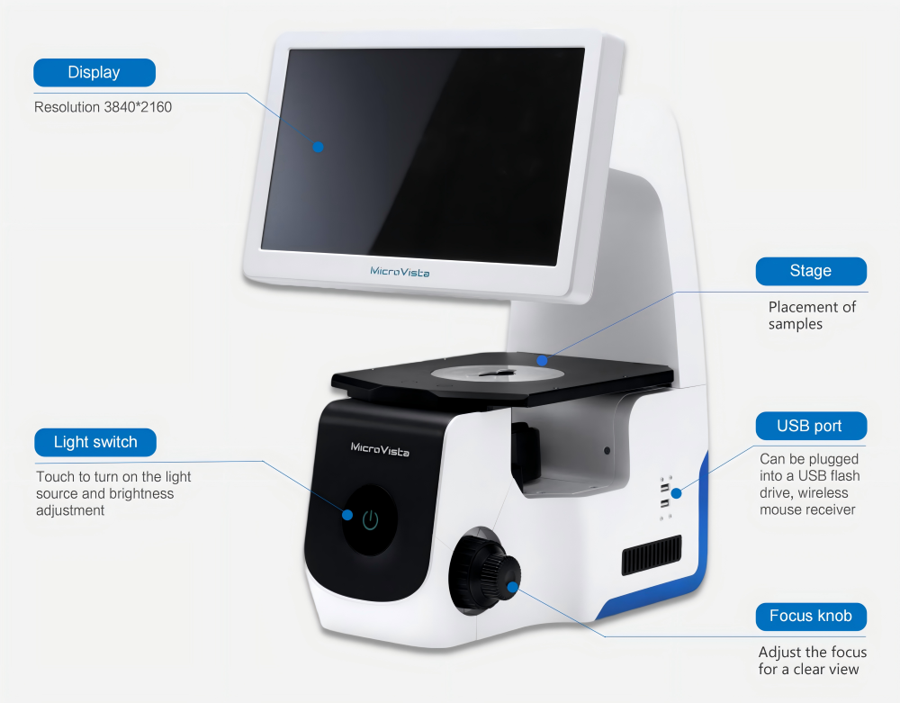
As an eyepiece-free microscope, Vita-X1 features a 4K digital tablet display with a resolution of up to 3840*2160 which is 4 times of traditional 1080P image, resulting in smooth visualization, as well as fine structure images with rich colors, high contrast, and smooth images.
technical specifications
illuminant | LED light source, up to 20,000 hours of life, touch intensity control | image acquisition | Touch control/mouse control |
Imaging mode | Bright field, contrast | software | Lunix embedded system, no need to connect to PC, has image/video acquisition, white balance, objective lens calibration, measurement and other functions |
collecting mirror | Numerical aperture 0.3, working distance 72mm | SC | 512G USB drive (randomly standard) |
rotating nosepiece | Four holes | data transmission | USB or HDMI connection |
Preloaded objective lens | 4x (NA 0.13),10x (NA 0.25),20x (NA 0.4)相差物镜. | Image format | TIFF/JPG |
objective table | Size: 252mm*200mm | weight | 9.64KG |
Tuning mechanism | Trip length: 20mm | size | 329.60mmX429mmX348mm |
image taking speed | 30 frames per second @4K resolution | Display size | 13.3 inches |
display resolution | 3840*2160 | adapter | Universal adapter, slide adapter, 35mm/60mm culture dish adapter |
objective (optional) | Long working distance difference objective 40x (NA 0.55), flat field achromatic and above | objective table Mobile Suite (optional) | The right handle flatbed is equipped with a removable multi-well plate specimen holder Platform travel: X=110mm, Y=74mm |
*For scientific use only