STEDYCON
STEDYCON is a newly designed ultra-high resolution microscope that can easily upgrade the four channel confocal (405nm, 488nm, 561nm, 640nm) STED (775nm) ultra-high resolution system on the basis of traditional wide field fluorescence microscopes! At the same time, it occupies a small space, is easy to use, and users can quickly achieve the acquisition of<40nm>
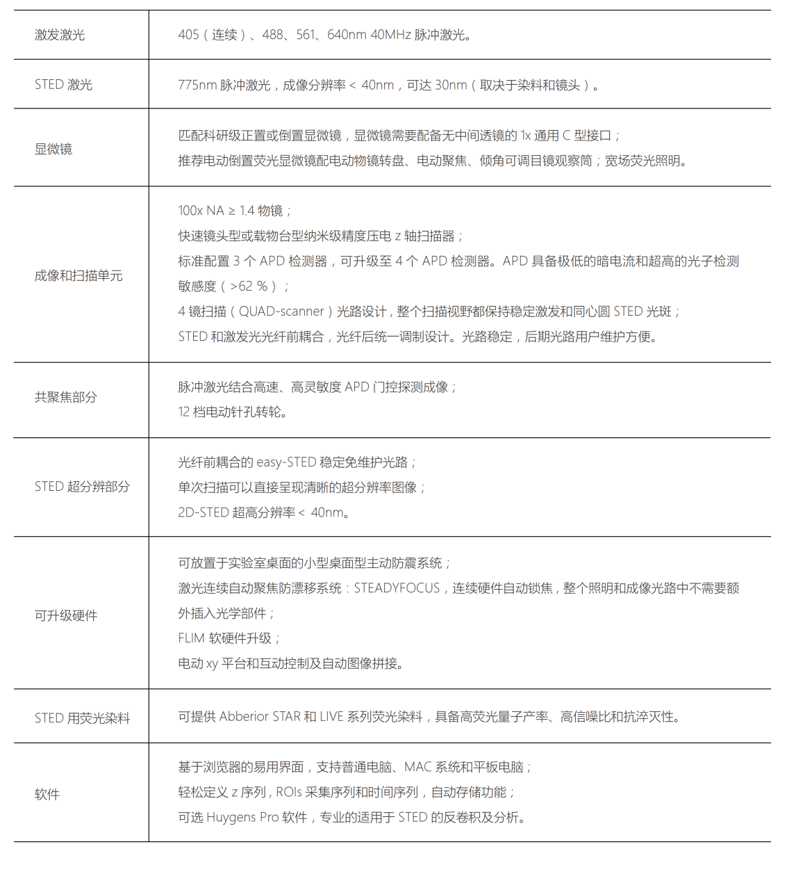
Comparison of confocal and STED imaging of two nuclear pore complex proteins: red gp210 (Abbey STAR580) and green PAN4/5 (Abbey STAR635P).
Note that gp210 is located in eight symmetrical groups around the nuclear pore complex.
Main features of the product
Highest quality confocal and STED effects
STED resolution<40nm, up="" to="" 30nm="">
High sensitivity detection system
All use high-sensitivity, high-speed, single photon counting APD (avalanche diode) gate controlled detectors;
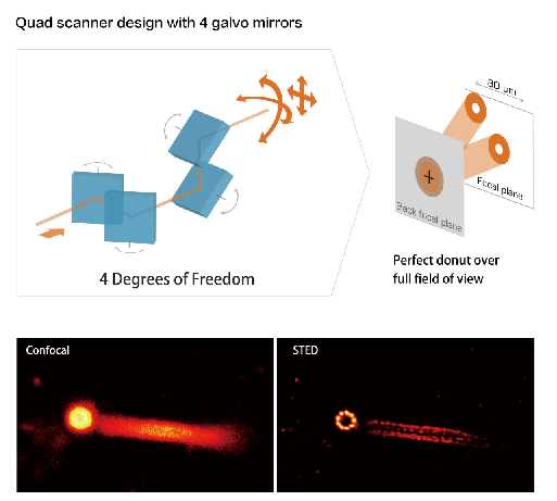
High precision 4-mirror scanning system
Ensure the quality of the super-resolution imaging spot under a large imaging field of view (80x80 μ m @ 100x), with a scanning resolution of 12000x12000;
Match research grade upright and inverted microscopes of major brands
Can quickly upgrade existing fluorescence microscopes to multi-color confocal microscopy and STED;
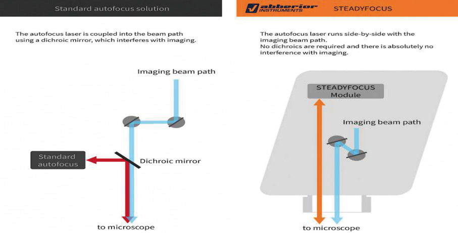
Ultra-stable
The all-new STEADYFOCUS module ensures that your images remain in precise focus for several days;
Large upgrade space
Easily achieve ultra-high resolution large field of view stitching imaging and ultra-high resolution fluorescence lifetime imaging (FLlM);
Space saving
The volume of the scanning head is only equivalent to a slightly larger CCD;
Plug and play
STEDYCON can be installed in just a few minutes, and super-resolution acquisition can be achieved with simple optical calibration;
Easy after-sales maintenance
The light beam is accurately coupled with the excitation laser before entering the fiber optic cable ("easySTED" optical calibration mode), and the optical path maintenance is simple;
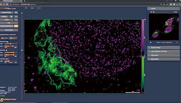
Browser based software interface
Flexible parameter control software, easy to learn and use; Supports Windows systems, MAC systems, and tablets.
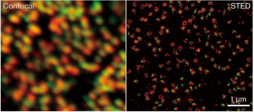
| Live cell mitochondrial inner membrane, microfilament STEDYCON imaging. Blue: PKMO of mitochondrial inner membrane; Red: Microfilaments SPY650 FastAct; Gray: Cell nucleus, SPY505-DNA Image source: Peking University, Dr. Tianyan Liu, Professor Zhixing Chen's laboratory |
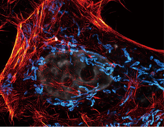
 | Comparison of endoplasmic reticulum confocal microscopy and STEDYCON imaging in live cells expressing novel fluorescent proteins Image source: Professor Fu Zhifei's laboratory, Fujian Medical University |
4-mirror beam scanning system
1)No scanning lens design, reducing beam deformation and loss; 2)High speed laser switch for precise control of scanning conversion angle laser switch, protecting samples; 3)STEDYCON adopts a scanning resolution of 12000 x12000, which can achieve fine resolution acquisition in a large field of view. |
Imaging of centrioles in fruit fly testes Image source: China Agricultural University, Dr. Zhang Jiawen, Professor Fu Jingyan's laboratory |
Pulse laser (STED and visible light excitation laser) combined with APD gating detection
1)可见光激发光和STED 光均使用精准耦合的脉冲激光,只需连续激光 1/12 的能量,就能达到与其相同的分辨率。Both visible light excitation light and STED light use precisely coupled pulsed lasers, requiring only 1/12 of the energy of continuous laser to achieve the same resolution. 2)High sensitivity APD (quantum efficiency>62% @ 680nm) was used for high-speed gating detection, and APD was used to match pulse excitation light and pulse STED light with high accuracy on the time axis during the acquisition time window, further improving the resolution and signal-to-noise ratio of the image. | 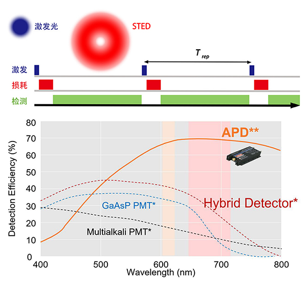 |
Easy calibration settings Easy-STED
The crucial guarantee for the high-resolution imaging quality of STED is the perfect alignment of STED pulsed laser and excitation light. Traditional STED microscopes use the method of modulating STED light first and then coupling it, which is extremely sensitive to the environment. Small environmental changes (such as temperature) can cause the spot to shift in the center, resulting in a significant decrease in STED quality and sometimes even worse results than confocal microscopy. Accurate spot tuning is very difficult for users without a deep optical background.
Abberior Instruments has developed a pre aligned Easy STED system based on new technology from the Stefan Hell team. The excitation light and STED beams are aligned before entering the fiber, and coaxial spot tuning is achieved in the rear optical path. After the spot is pre-set, the entire optical path is almost unaffected by any environmental changes, thus solving the problem of repeated calibration of dual beams and allowing users to focus on the experiment itself and imaging operations. | 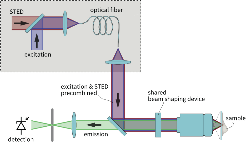 |
Easy to operate STED software
STEDYCON product is an easy-to-use super-resolution microscope. Whether novice or expert users, clear STED ultra-high resolution images can be obtained through a single user interface and a few clicks, and different fluorescence ultra-high resolution images and confocal images can be directly integrated into one interface. Such a concise and professional operation process provides users with a good operating experience.
STEADYFOCUS Fully automatic hardware focus locking module
The new one click STEADYFOCUS fully automatic coke locking module is a continuous hardware coke locking module specially designed by the team for STEDYCON. It ensures no focal drift in confocal and STED imaging during several days of experiments. Unlike other typical hardware anti drift designs, STEADYFOCUS does not require additional optical components to be inserted into the microscope imaging beam, so there is no fluorescence loss or image distortion. This automatic focusing module supports multiple media objectives (water, oil, silicone oil, glycerin) and sealing media (water, Mowiol, etc.) as well as mainstream inverted microscope bodies. STEADYFOCUS uses a piezoelectric focusing device that does not rely on the electric Z-axis of the microscope, and can even achieve automatic focusing function on manual microscopes. | 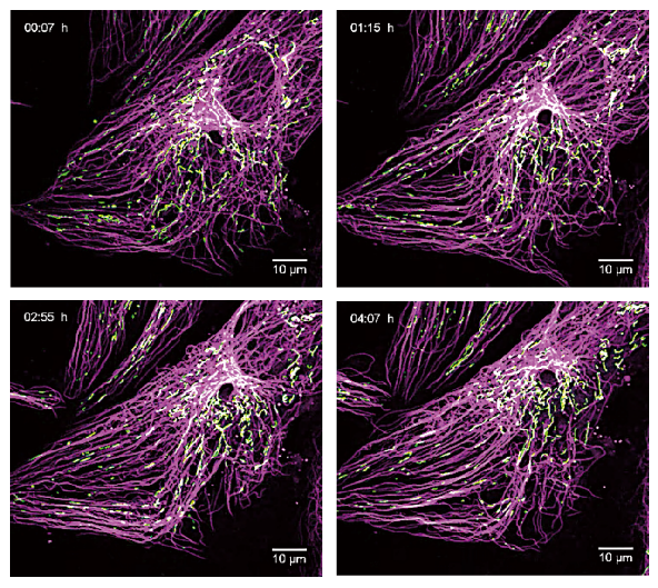
Multi color live cell confocal time-lapse imaging using STEADYFOCUS for up to 13 hours |
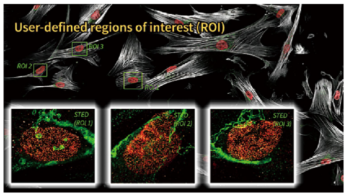
Ultra high resolution stitching multi position imaging
STEDYCON can control the electric XY stage to automatically scan the entire millimeter sized sample in a large field of view. The software can control interactive navigation and imaging throughout the entire field of view of the sample. Multiple regions of interest (ROIs) can be set, and automatic image recording and stitching of multiple ROIs can be performed sequentially using confocal or STED (this feature requires additional purchase of Huygens plugin).
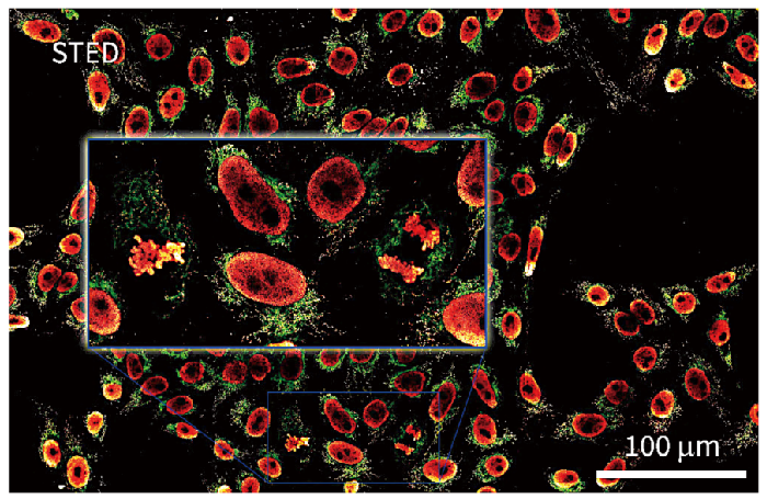 | Large area super-resolution STED imaging. Ultra high resolution stitched multi position imaging helps capture rare events such as cell division. Sample: Mammalian cell immune marker mitochondrial protein TOM20 (Abbey STAR Red, red) and double stranded DNA antibody to display mitochondrial DNA (Abbey STAR Orange, green) |
 | The sample is mammalian cells Red represents the nuclear pore complex, green represents the Golgi apparatus |
Easily upgrade to a super-resolution time-domain fluorescence lifetime imaging (FDM/FCS) system with the following main functions

You can directly use the pulse laser and single photon counting APD detector configured by the system itself without upgrading these hardware components | 
STED and FLIM/FCS dual system synchronous raw data sharing and result display | 
High precision real-time scientific fitting calculation FLIM/FCS or offline calculation | 
TCSPC hardware and analysis software based on PicoQuant or Becker&Hickle |
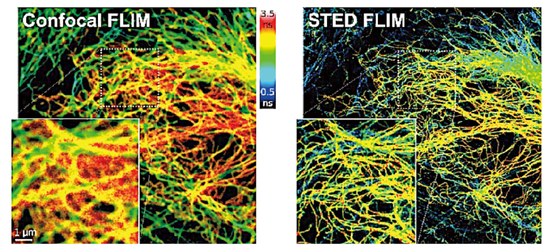
Fluorescent labeled microtubule protein (Atto647N) and vimentin protein (Abbey STAR 635P)
 | Green: Tubulin marked with Abbey STAR Red, Red: Clathrin marked with Abbey STAR Orange Image source: from the laboratory of Dr. Hao Huiwen and Professor Sun Yujie at Peking University |
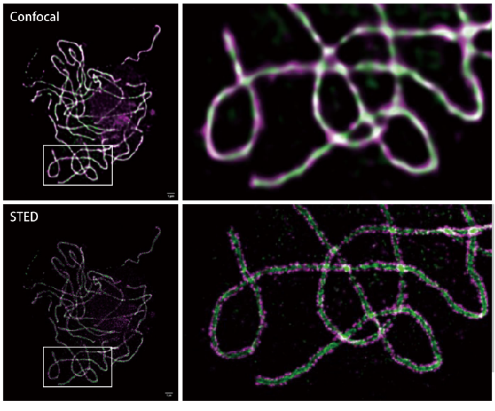 | Comparison of confocal microscopy and STEDYCON imaging of DNA synapse complexes in rice pollen mother cells Green Zep1, Purple Rec8 Image source: Institute of Genetics and Development, Chinese Academy of Sciences, Dr. Miao Yongjie, Professor Cheng Zhukuan's laboratory |
Product parameters