Products and Services
Home > Products and Services
Light-Sheet Price:¥

A mild, fast, and versatile single objective optical microscope.
· Zero hands-on cost
· Mild long-term imaging
· High axial resolution reveals the mysteries of real three-dimensional space
· ​From live cells, organoids to whole organs

Online inquiry
Detailed

Subverting tradition

Redefine the Light Sheet

The latest generation of single objective 3D optical imaging technology, which combines illumination detection with a single objective lens, saves you the need for complex system optical path adjustment and greatly enriches your sample operation space.


32f6ae07-a951-45bc-8ea3-5dc2e943d924.jpg

ea47e344-7796-4105-ad23-12258c7fab2b.jpg

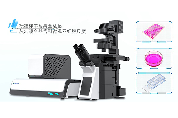
Universal light film for all scenes

From glass slides, culture dishes to well plates

Thanks to its unique single objective lens mode, SmartView is suitable for various application scenarios. The open top optical design allows you to achieve one click 3D imaging with simple sample loading operations. At the same time, due to compatibility with electric switching at different magnifications, in situ cultivation and observation of samples can be achieved.


Realistic display

3D spatial details

The unique multi-level diffraction free light field control technology can generate the thinnest "450 nm" optical knife, achieving ultra-high tomographic accuracy.


a1f182c9-8f7d-4a32-ad8d-296bb47d7edf.jpg

Optical characteristics

Reduce phototoxicity and photobleaching

Laser tomography illumination technology achieves selective planar illumination of samples, greatly reducing the system's phototoxicity to samples and facilitating non-invasive long-term dynamic detection of biological samples. At the same time, without the need for tilted plane correction in optical microscopy design, the utilization efficiency of fluorescence signals can be greatly improved.


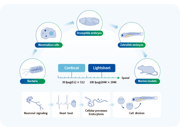
From live cells, organoids to whole organs

The multimodal optical microscopy imaging system can achieve seamless switching between bright field, wide field, and optical sheet in situ. The SmartView with inverted single objective mode design allows for a diverse range of research objects, meeting your needs for imaging speed, resolution, phototoxicity, depth, and more.


1c292988-64b7-4e10-ae06-b585de966d6c.jpg

726e333c-ff52-4091-8529-a7d7806e37a4.jpg

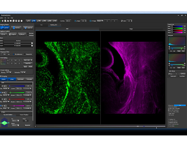
Intelligent image acquisition software

Our self-developed high-throughput image acquisition software supports intelligent multi-dimensional image acquisition. It can achieve user-defined imaging modes such as multi-color imaging, time series imaging, multi-point imaging, z-axis tomography imaging, and large image stitching.


AI Empowerment

The self supervised AI image signal-to-noise ratio enhancement algorithm can perform high fidelity image enhancement on weak fluorescence signals without the need for labeled data. The related technology has been published in Nature Communications.
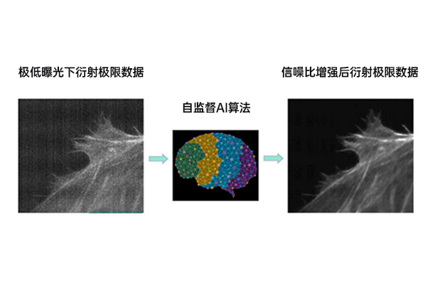

31fc3efc-cece-49b8-9dbe-8bf7ddb11a54.jpg

U2OS cells

Live cell time series imaging was performed using a 100 x/N.A.1.5 oil microscope to capture 200 time points for a total duration of 1 hour.

U20S cells

Time series imaging of EGFP labeled EB3 protein using a 100 x/N.A.15 oil microscope. Zdepth coding display.

Zebrafish

24 hpf dual color 3D imaging of zebrafish. 27 fields of view were captured using a 30 x/N.A1.05 silicone oil objective lens for large image stitching. Each field of view captured 582 layers at a step size of 0.65 um/slice, with a total imaging time of 52 minutes. Green represents the nucleus, while purple represents the extracellular vesicles. The sample was provided by Professor Yuan Zhengwei's team from Shengjing Hospital affiliated with China Medical University.

Fruit fly (head)

Transparent processing of three-dimensional imaging of adult fruit fly heads. We used a 30 x/N.A.1.05 oil mirror to stitch together 30 large images of 5 * 6. A total of 874 layers were collected at a step size of 0.54 um/slice for each field of view, with a total imaging time of 65 minutes. The sample was provided by the team of Professor Zhang Yongqing from Hubei University.

Mouse spleen

Mouse spleen samples captured using a 10x/N.A. 0.4x microscope, with vascular markers. The sample size is 4.8 mm x 9.5 mm x 2.7 mm. 2054 layers were captured with a step size of 1.3 um/slice and an exposure time of 400 ms.

Function

Parameter

Excitation wavelength

405 nm, 488 nm, 532 nm, 561 nm, 589 nm, 640 nm (flexible choice)

Spatial resolution

220 nm x 220 nm x 450 nm @100 x obj; (AI super resolution reconstruction resolution can reach 100nm x 100nm x 100nm, optional)

Time resolution

100 fps @2048 × 2048

Detect the spectral range

400 nm - 740 nm

Thickness of the light sheet

*** Thin 450 nm, thickness adjustable

Image depth

3 mm

Long-term observations

Hours to days

Post-image algorithm

AI image enhancement; AI intelligent image analysis

Visual field

130 μm × 130 μm @100 × obj; 3.3 mm × 3.3 mm @4 × obj


© 2026 Guangwei Jing Biological Technology (Shanghai) Co., Ltd.  All Rights Reserved.